
Hepatotoxicity and ADME services


Why us
Our NANOSTACKSTM-based in vitro models recapitulate the cell-cell crosstalk, fluid flow and spatial cellular arrangement typical of the in vivo environment. This way, toxicity screening on our models can yield more accurate results than the use of traditional models. We provide in vitro testing services using the in vitro model of your choice.
Why choose our NANOSTACKSTM liver models
Superior performance compared to other hepatic models
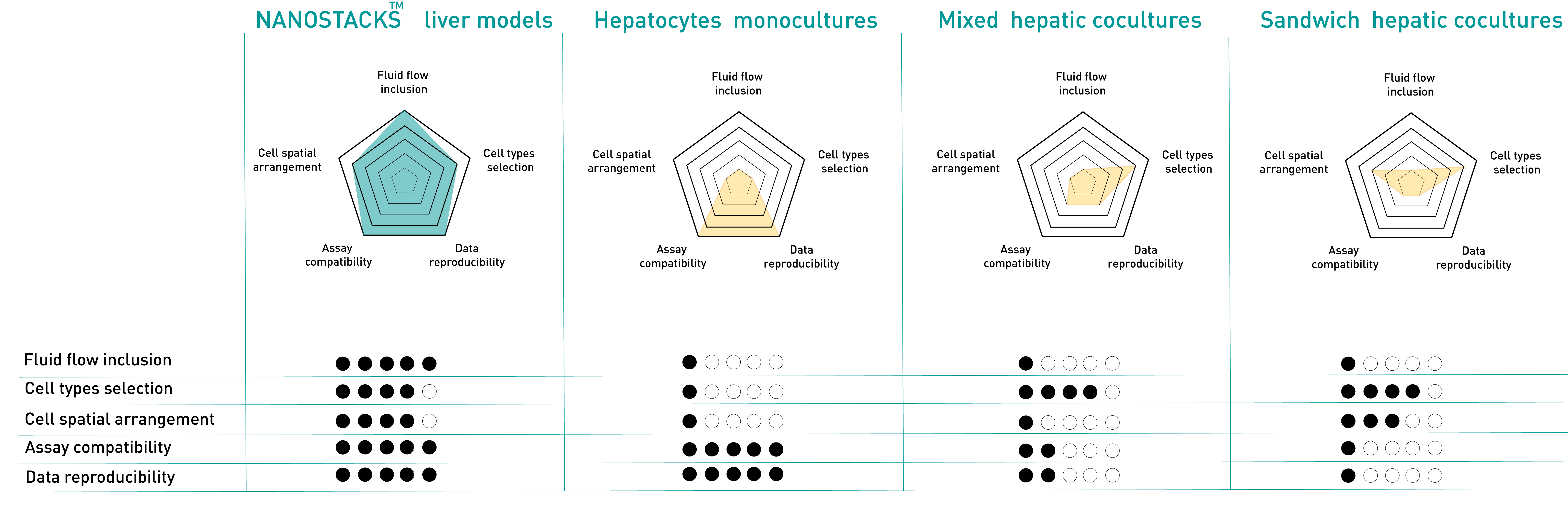
Click on the options below to explore our validated solutions for hepatotoxicity and ADME studies
NANOSTACKSTM liver monoculture model
NANOSTACKSTM liver triculture model
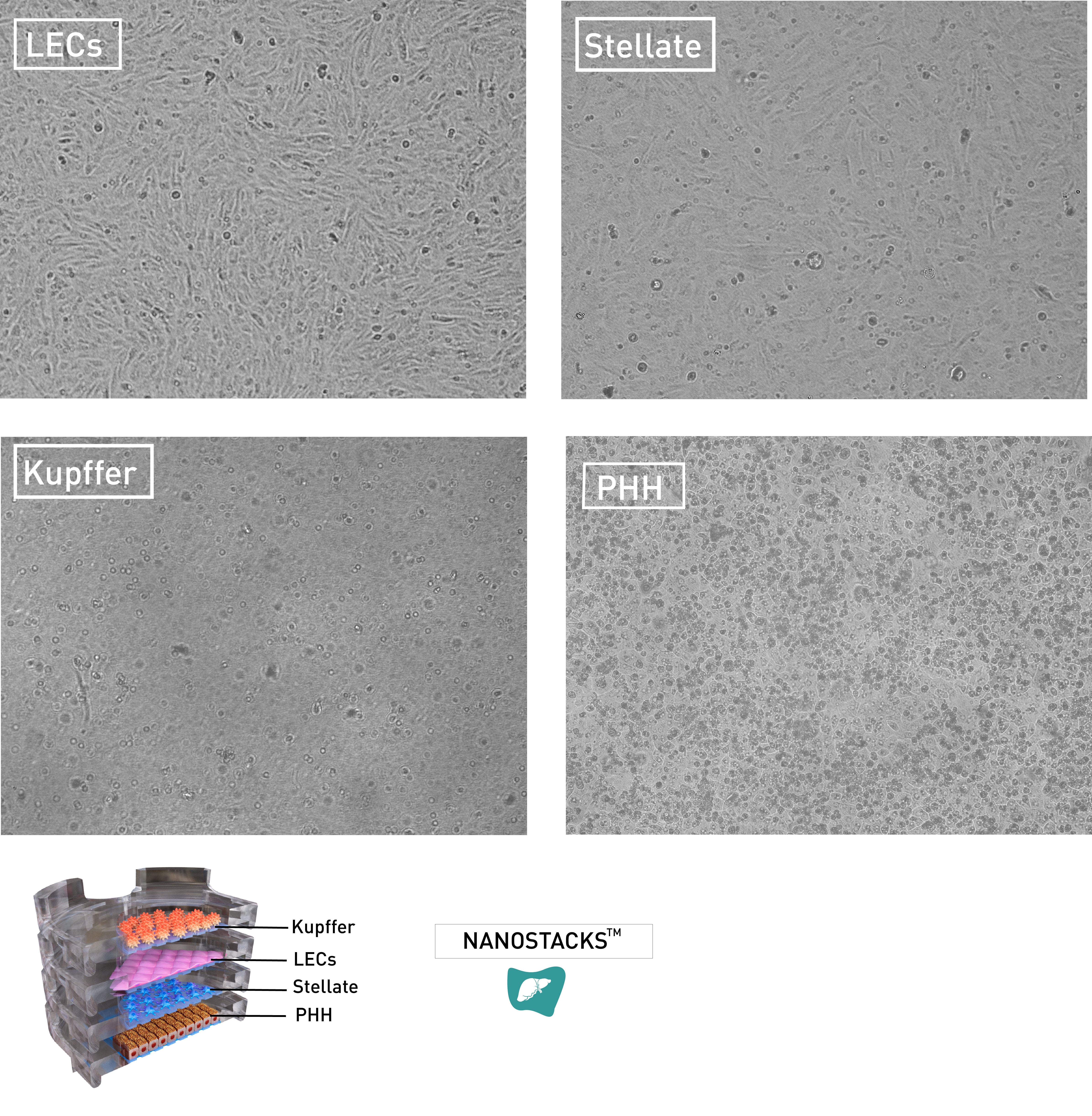
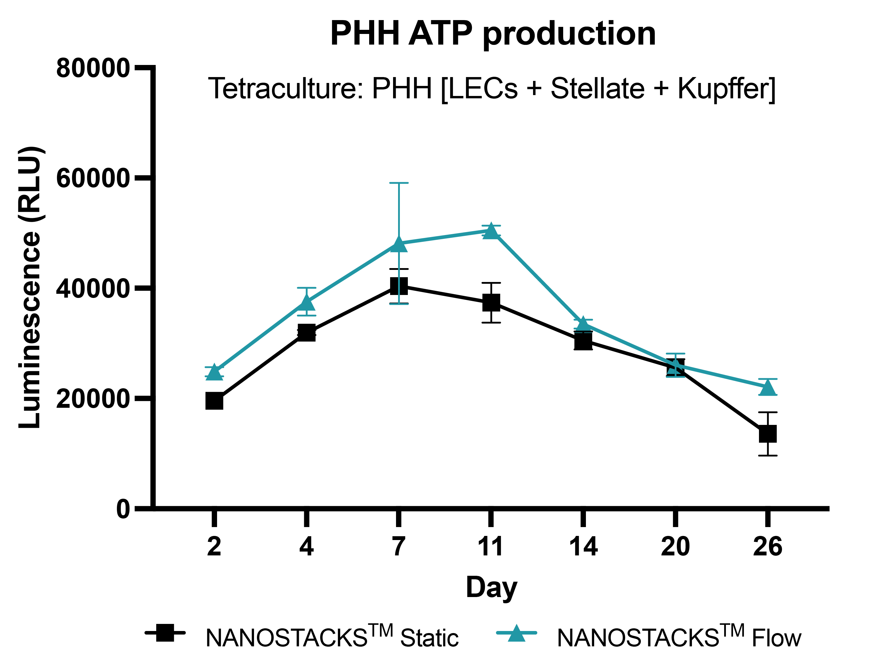
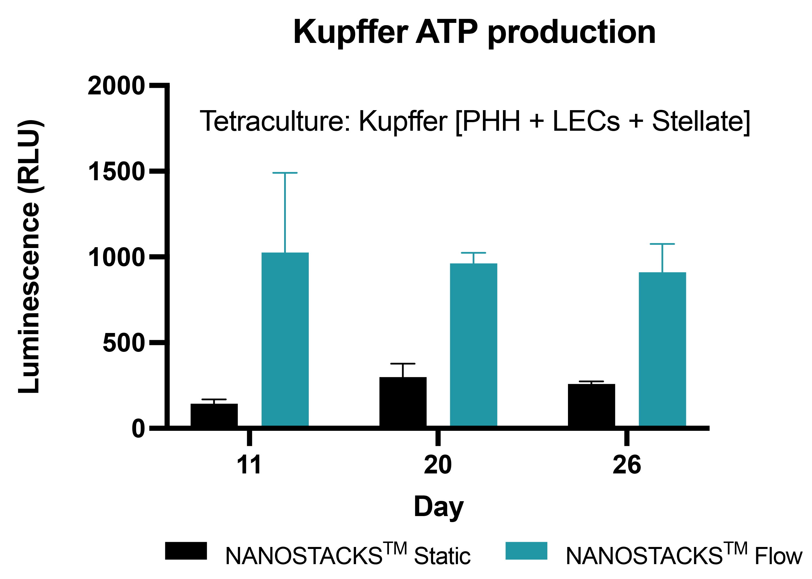
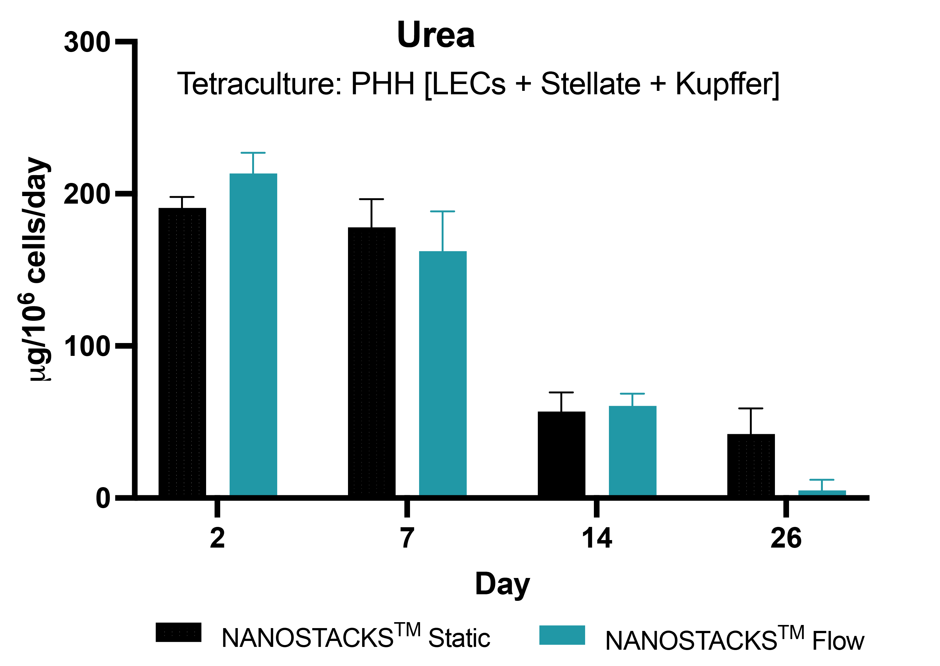
NANOSTACKSTM liver tetraculture model

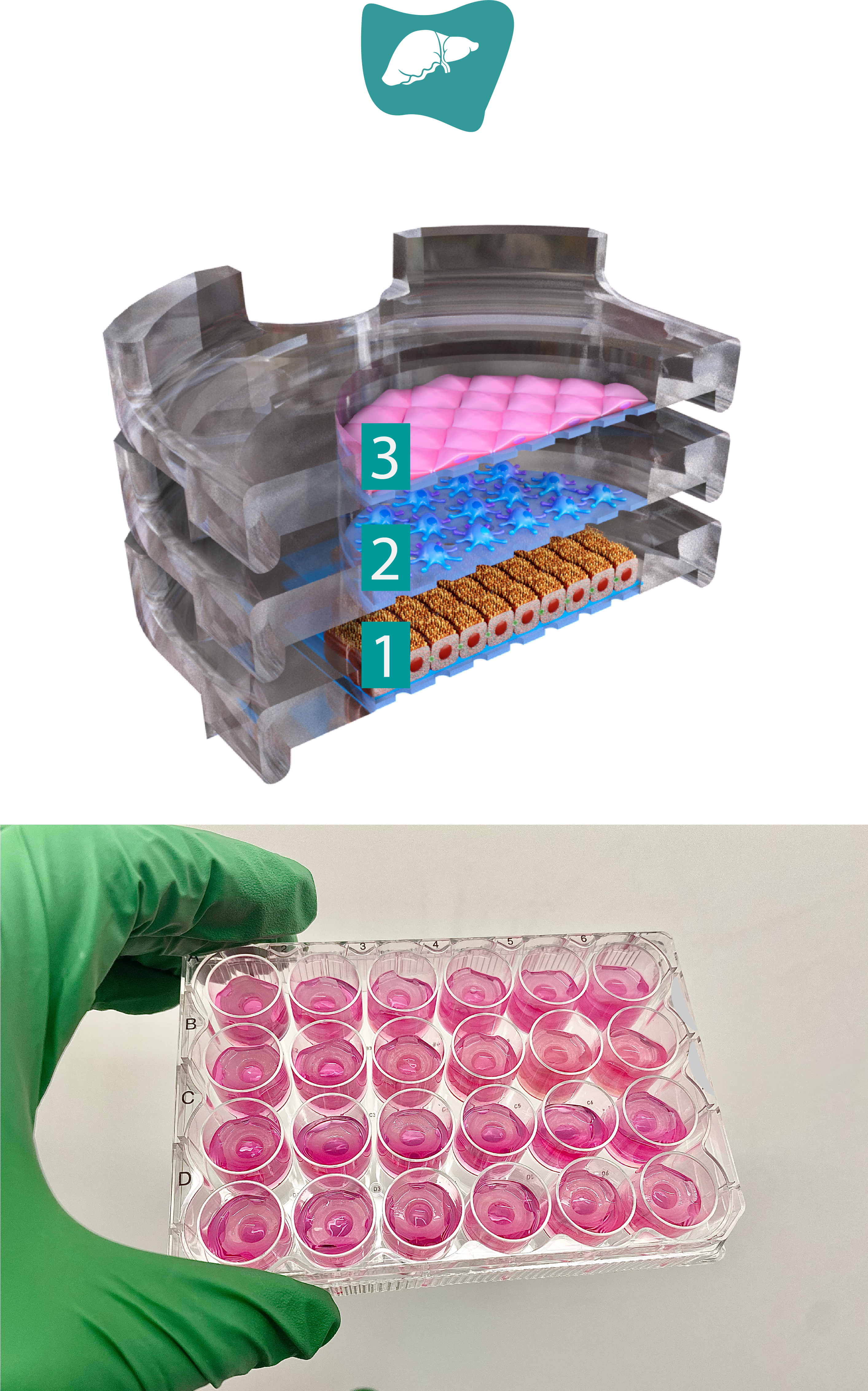
The model is built using our proprietary NANOSTACKS™ technology
- Layer 1: Hepatocytes compartment
- Cell type: Primary human hepatocytes
- Primary human hepatocytes (PHH) are the gold standard for hepatotoxicity screening due to their physiological relevance and ability to recapitulate key liver functions, such as CYP activity and albumin production. The model can be utilized in various ways, including as a standalone model or as a control for our triculture and tetraculture models.
- Fluid flow included
OUTPUTS
Contact us for alternative endpoints.
Comprehensively validated liver monoculture model

Liver PHH cells on NANOSTACKSTM
Representative widefield images of primary human hepatocytes (PHH) in NANOSTACKSTM. Magnification: 10X.

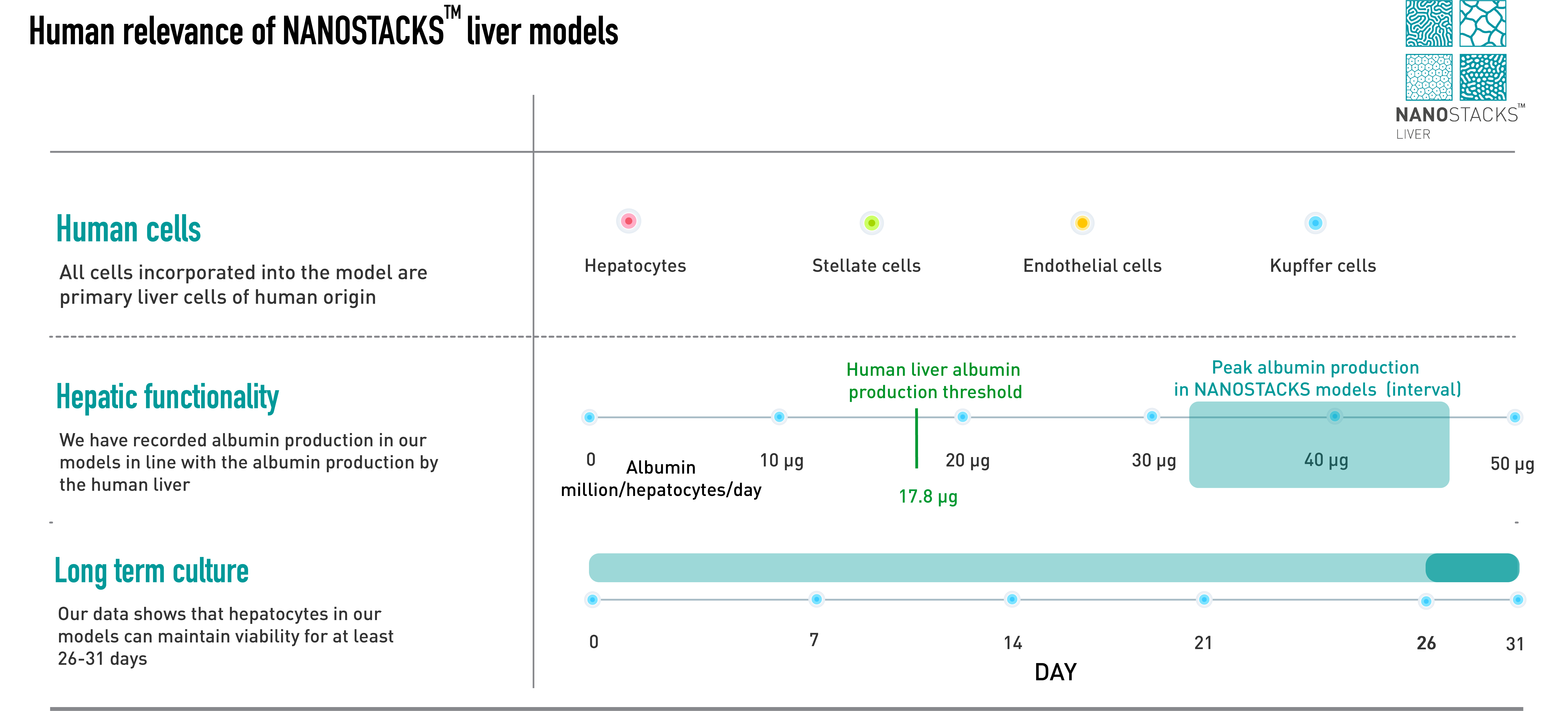
Long term model culture
Primary human hepatocytes (PHH) grown on NANOSTACKSTM under both flow and static conditions maintained viability for 31 days of the study, with no substantial differences between static and flow conditions. Data: mean ± SEM. N = 3 on day 2, 4, 7,11, 14, 20 and 26.
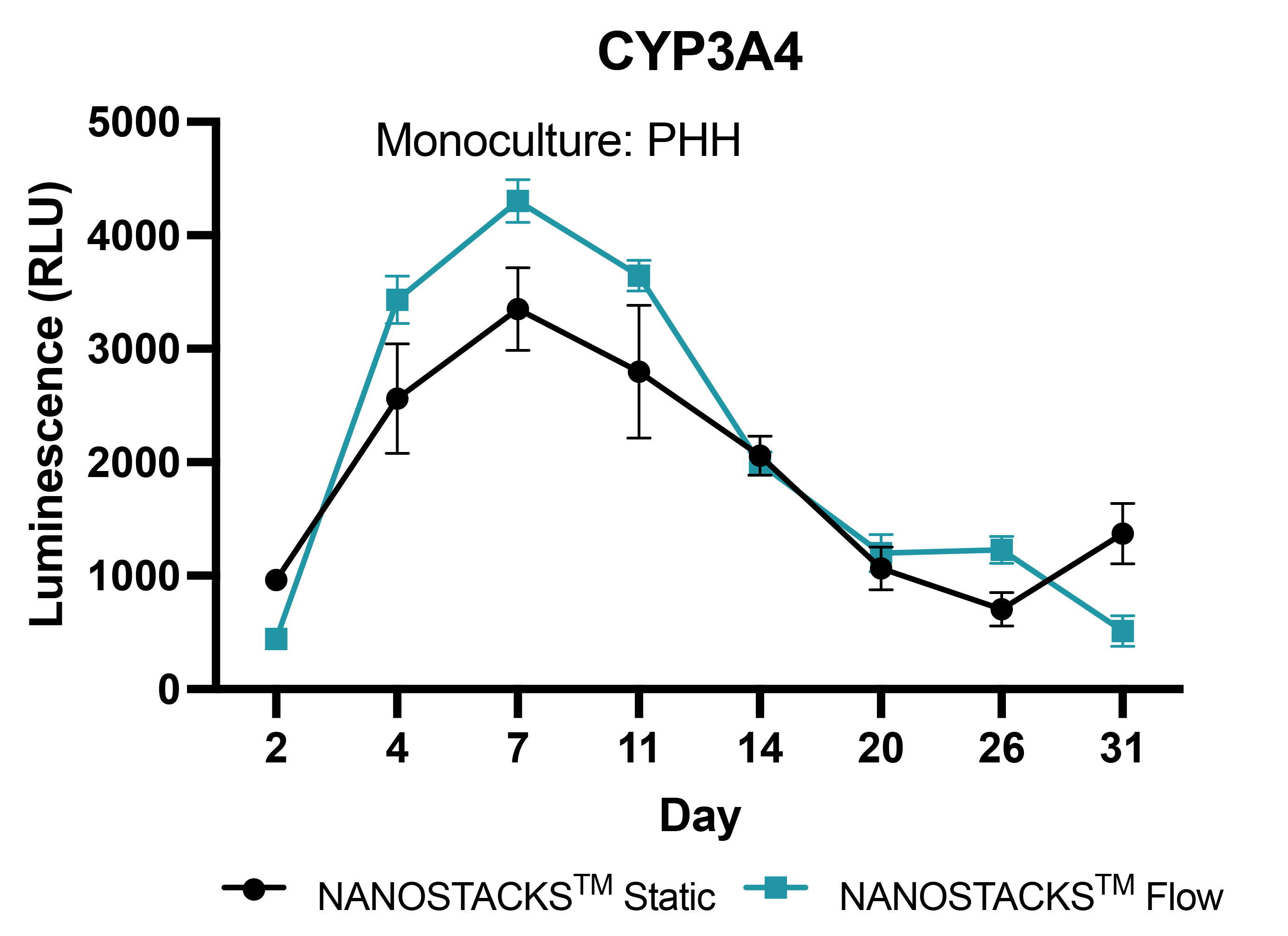
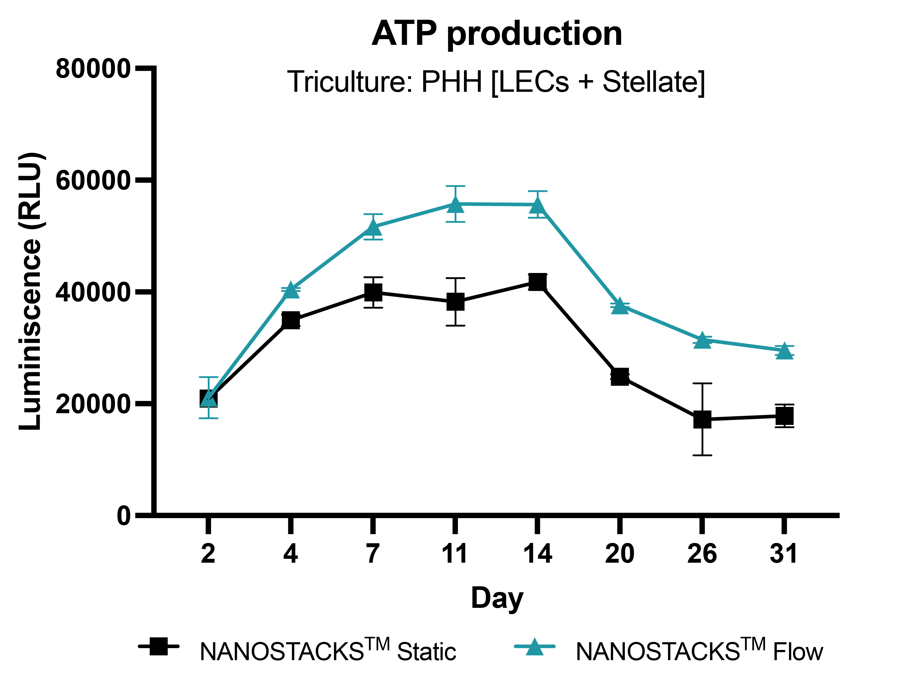
Sustained metabolic competence
The NANOSTACKSTM model of primary hepatocytes (PHH) maintained active metabolic activity throughout the 31-day study period. However, the introduction of flow into the model resulted in significantly higher CYP3A4 activity between days 4 and 14 compared to the static model. Data: mean ± SEM, N = 3 on days 2, 4, 7, 11, 14, 20, 26, and 31.

Human-like albumin levels
Albumin production in primary human hepatocytes reached human threshold levels (17.8 µg/million/hepatocytes) by day 4 of culture, peaking at approximately 40 µg on day 7. The model maintained above threshold levels until day 26 in both static and flow conditions, except on day 26 when albumin levels in the flow condition decreased to 8.9 µg. Data: mean ± SEM. N = 3 on days 2, 4, 7, 11, 14, 20, and 26.
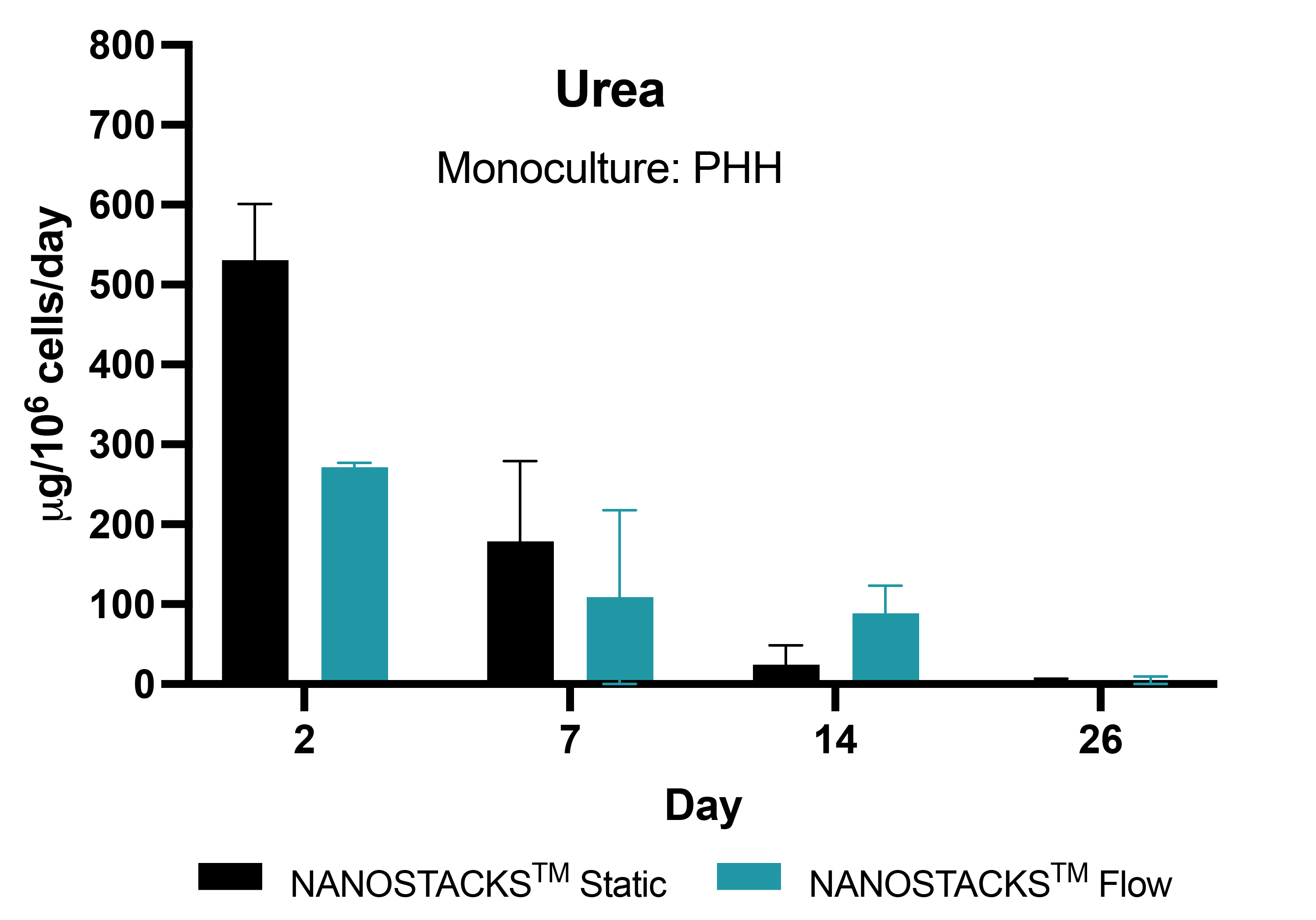
Human-like urea levels
Under static conditions, human primary hepatocytes (PHH) on NANOSTACKSTM maintained human-relevant levels of urea (>56 µg/million/hepatocytes/day) for 7 days, decreasing to 24.25 µg by day 14. Conversely, when flow was introduced into the model, the human urea level threshold was extended until day 14. Data: mean ± SEM, N = 3 on days 2, 4, 14 and 26.

Clinical translatability
The NANOSTACKSTM human primary hepatocytes (PHH) model accurately predicts hepatotoxicity of Zileuton, an FDA-classified high-DILI-concern drug, at clinically relevant concentrations following 6 days of repeated exposure, demonstrating its robust DILI prediction capability. Data: mean ± SEM. N = 3.
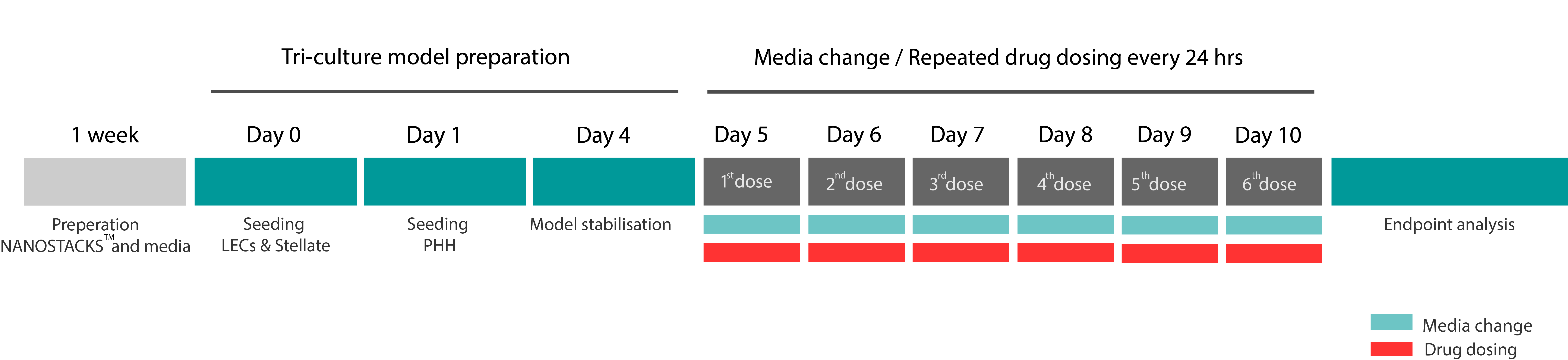
PHH monoculture model DILI screening standard process & timeline*

*Dosing intervals and duration can be adjusted based on your requirements


Unique Benefits
Human Relevance
Developed entirely using primary human cells, ensuring translational value with consistent results.

Intercellular communication
Cell-cell crosstalk between key hepatic cells is replicated in the model, increasing its similarity to the in vivo environment.
Inclusion of fluid flow
Fluid flow can be included in the model, modelling the natural mechanical environment of the liver.
Differential toxicity evaluation
Toxicity can be evaluated on each individual cell type included in the model.
Long-term studies
Cells included the model are viable up to 31 days, providing the possibility to perform long-term toxicity screenings.
High quality cells
Our cells are obtained from trusted suppliers and consistently perform key hepatic functions.
Get in Touch
Explore how our models support your research studies

Alternative solutions

MODEL SHIPPING
Alternatively to performing our services within our facilities, we can also ship our validated organ models directly to you lab ready for immediate use.
CUSTOM MODEL DEVELOPMENT
We can develop custom organ models using our NANOSTACKSTM 'Plug&Play' technology based on your requirements.
Need some help?
If you're interested in discovering how our organ models can benefit your research, we invite you to get in touch with us. Our team is available to provide further insights and address any inquiries you may have.